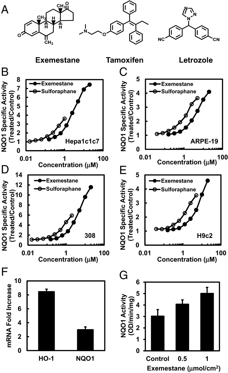
Fig. 1.
Exemestane induces cytoprotective enzymes in cultured cells and mouse skin. (A) Chemical structures of exemestane, tamoxifen, and letrozole. (B–E) Dose-dependent induction of NAD(P)H:quinone oxidoreductase 1 (NQO1) by exemestane and sulforaphane in different cell types: Hepa1c1c7, ARPE-19, 308 keratinocytes, and H9c2 cardiomyocytes. Cells were plated in 96-well plates, and 24 h later were exposed to serial dilutions of exemestane or sulforaphane for a further 48 h. NQO1 activity is expressed as mean ratios of treated over control specific activities by using eight replicate wells for each inducer concentration. SDs for all measurements were less than 10%. (F) Real-time PCR analysis of phase 2-related genes heme oxygenase-1 (HO-1) and NQO1 expression after 6 h of treatment with exemestane in H9c2 cells. β-Actin was used as an endogenous control for the target genes, and values are represented as the ratio of change in the mRNA levels of exemestane-treated versus vehicle-treated cells. Means ± SD are shown. (G) Induction of NQO1 in mouse skin by topical application of exemestane. The back of each SKH-1 hairless mouse (n = 5) was topically treated with two concentrations of exemestane [0.5 and 1.0 μmol/cm2 in 80% (vol/vol) acetone] and solvent only over about a 2.0-cm2 area for three doses at 24-h intervals. Mice were euthanized 24 h after the last dose, and dorsal skin was harvested. NQO1 specific activity was measured in supernatant fractions of homogenates of skin sections treated with exemestane or solvent (control). Means ± SD are shown.

