
Fig. 2.
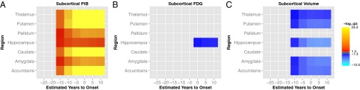
Comparison of [11C]Pittsburgh compound B (PiB) (A), FDG (B), and volume (C) between carriers and noncarriers in subcortical gray matter. Significant increases (FDR corrected P < 0.05) are indicated in red/orange and decreases in blue/cyan. Elevated PiB uptake was detected by EYO = −15 in all of the subcortical gray matter structures. Excluding the pallidum and caudate, all subcortical structures exhibited volumetric decline ten years before the expected age of onset. Metabolic decreases were only seen in the hippocampus, and only after the estimated onset of symptoms.