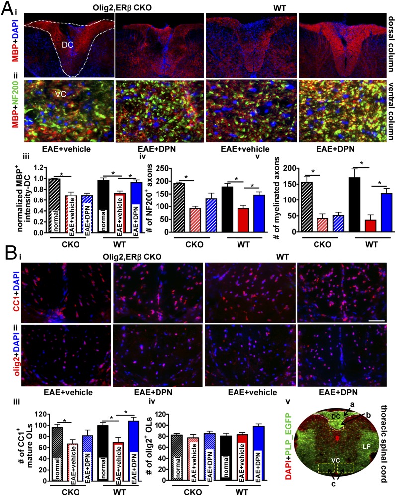
Fig. 4.
Selective deletion of ERβ in OLs prevents DPN-induced improvement in myelin density and mature OL numbers, but partially protects against axonal loss within the spinal cord of EAE mice. Dorsal column of thoracic spinal cord sections from Olig2,ERβ CKO and WT EAE mice were examined for myelin density (A, i; red). Quantification reveals improved MBP+ intensity in DPN-treated WT EAE but not DPN-treated Olig2,ERβ CKO EAE mice (A, iii). Ventral column of thoracic spinal cord sections were imaged at ×40 and depict costain using NF200 (green) and MBP (red; A, ii). Quantification reveals improved NF200+ axon numbers upon treatment with DPN in WT animals and a trend toward this effect in Olig2,ERβ CKO mice (A, iv). Increased numbers of MBP+ and NF200+ axons in DPN-treated WT, but not Olig2,ERβ CKO, EAE mice (A, v) was observed. For both genotypic cohorts, vehicle-treated mice displayed a significant reduction in myelinated axon numbers (normal; A, iii–v). Vehicle-treated Olig2,ERβ CKO and WT animals exhibited a reduction in CC1+ OLs (B, iii). DPN treatment in WT, but not Olig2,ERβ CKO, EAE mice improved mature OL numbers (B, iii). Olig2+ cell numbers were similar across all groups (B, iv). PLP_EGFP normal thoracic spinal cord sections (×4 magnification) were costained with DAPI (red). White square boxes and a, b, and c denote various areas of the spinal cord used for quantification (DC, dorsal column; LF, lateral funiculus; VC, ventral column; B, v). *P < 0.05; ANOVAs; Bonferroni's multiple comparison posttest; n = 8–10 mice per treatment group.

