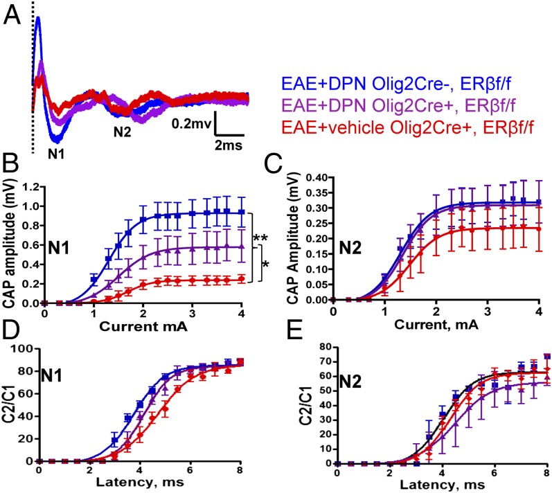
Fig. 5.
Selective deletion of ERβ in OLs prevents DPN-induced improvement in myelinated CC axon conduction. (A) Typical CAPs from various treatment groups at postinduction day 30 are shown. (B) An improvement in N1 CAP amplitude is evident in DPN-treated littermate control but not DPN-treated Olig2,ERβ CKO CC axons. (C) N2 CAP amplitude measurement reveals no difference. Average C2/C1 ratios were fitted to Boltzmann sigmoid curves (see ref. 1). (D) DPN-treated littermate control CC axons displayed a small but significant leftward shift. (E) No difference in N2 refractoriness was detected. n = 6. Statistically significant compared with normal controls at 1.1 ± 0.15 mA stimulus strength (*P < 0.05; **P < 0.001; ANOVAs; Bonferroni’s multiple comparisons posttest).

