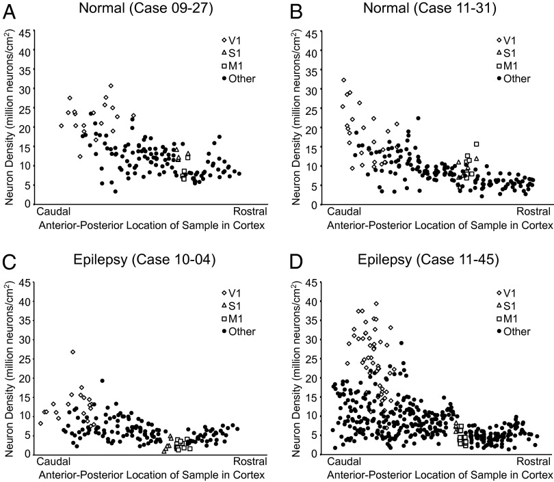
Fig. 2.
Neuron density versus the anterior-posterior dimension was plotted for each case. All flattened hemispheres were dissected into tissue pieces, and each piece was assigned an anterior-posterior coordinate by generating centroid measures. Normal neuron distribution (A and B) is shown to follow the caudal-to-rostral decrease in cortical neuron density that is typical of primates. Epileptic baboons show a reduction of neurons within this distribution, particularly in cortex rostral to the central sulcus (C and D).

