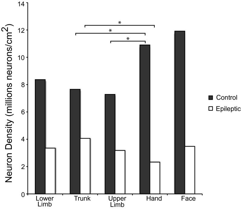
Fig. 3.
Histograms of neuron density in M1. Movement representation boundaries within M1 were estimated and dissected as described by Young et al. (16), and neuron densities by surface area were plotted for each case. There is neuron reduction within M1 of epileptic baboons relative to normal baboons. Lateral M1, which contains the face and hand representations, was the most neuron dense M1 region in normal baboons. In epileptic baboons, there is a substantial reduction of neurons in M1 in both cases, with the hand movement representation being the least neuron dense region of motor cortex.

