
Fig. 4.
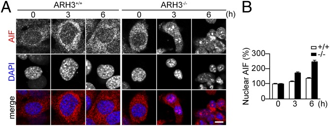
ARH3 deficiency enhanced AIF accumulation in the nucleus after H2O2 exposure. (A) AIF accumulation in nuclei of ARH3−/− MEFs after 3- or 6-h exposure to 300 μM H2O2. Cells were subjected to immunocytochemistry by using anti-AIF antibody (red in merged images) and DAPI staining (blue in merged images). (Scale bar: 10 μm.) These representative data have been replicated three times with similar results. (B) Mean AIF fluorescence in nuclei (means ± SEM, n = 36–40 cells).