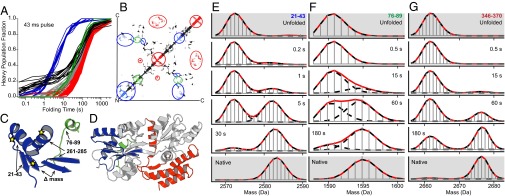
Fig. 4.

The folding history of different segments. (A) Blue and black segments are from Fig. 3B. Blue segments go on to form the 7-s intermediate, which persists through subsequent folding (E). Black curves track the early protected segments, which do not contribute to the 7-s intermediate. Segments that fold in the very slow broader grouping are graded from early (green), through gray, to late (red). (B) Contact map colored as for A. (C and D) Placement of the blue, green, and red segments in native MBP. (E–G) HX mass spectra comparing the time-dependent folding of some color-marked segments. The blue 7-s structure includes residues 9–43, 60–62, and 260–278. Residues 78–89 are shown in green; residues 180–195 and 315–370 are in red. Pulse time was 43 ms.
