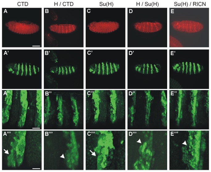
Figure 6. Nuclear localization of CTD and Su(H).
Either CTD or Su(H) were expressed in the presence or absence of either Hairless or RICN in a zebra pattern in the embryo using prd-Gal4 as indicated. The upper panel (A-D) shows Hairless protein in red except for (E), where the Notch intracellular domain was detected. The lower panels show expression of Su(H) and myc-CTD (green) as indicated. (A’’’-E’’’) are enlargements of (A’-E’’).
(A-A’’’) Whereas myc-CTD is mostly found in the cytoplasm (CTD, arrow in A’’’), it is predominantly nuclear when coexpressed with Hairless (H / CTD; arrowhead in B’’’). Su(H) is likewise nuclear and cytoplasmic upon overexpression (arrow in C’’’), however appears enriched in the nucleus (arrowhead in D’’’ and E’’’) when co-expressed with either Hairless (H / Su(H)) or RICN (Su(H) / RICN). Genotypes are: prd-Gal4 / UAS-myc-CTD / + (A-A’’’), prd-Gal4 / UAS-HFL UAS-myc-CTD / + (B-B’’’); prd-Gal4 / UAS-Su(H) / + (C-C’’’), prd-Gal4 / UAS-HFL UAS-Su(H) / + (D-D’’’), prd-Gal4 / +; UAS-Su(H) UAS-RICN/ + (E-E’’’). Size bars in A-E, A’-E’ 100 µm; in A’’-E’’ 25 µm; in A’’’-E’’’ 10 µm.

