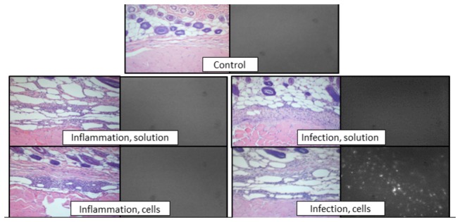
Figure 4. Punctuate fluorescence at the inoculation site of animals receiving injection of ICG-loaded cells.

Left image: representative paraffin-embedded sections from inoculation site or control limb stained with hematoxylin and eosin and imaged using light microscopy. Right image: representative frozen sections imaged with a near infrared filter.
