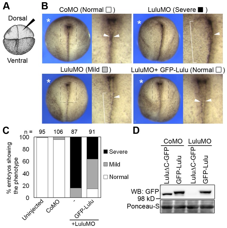
Figure 1. Lulu is required for neural plate hinge point formation.

(A) A four-cell stage embryo viewing from the animal pole. Injections were done in one dorsal animal blastomere (arrowhead). (B) Top views of stage 18 embryos unilaterally injected with indicated morpholinos (MOs, 20 ng) and GFP-Lulu mRNA (25 pg). Asterisks mark the injected side. Squared areas are magnified on the right of each panel. Arrowheads point to the hinge, visible as a pigment line, and brackets indicate weakening or disappearance of the hinge. For quantification of defects, lack of the pigment line was scored as “severe”, and weak or discontinuous pigment line was scored as “mild.” (C) Frequencies of defects shown in (B). (D) Lysates from embryos injected with indicated mRNAs and MOs were subjected to western blot. Ponceau-S staining shows loading.
