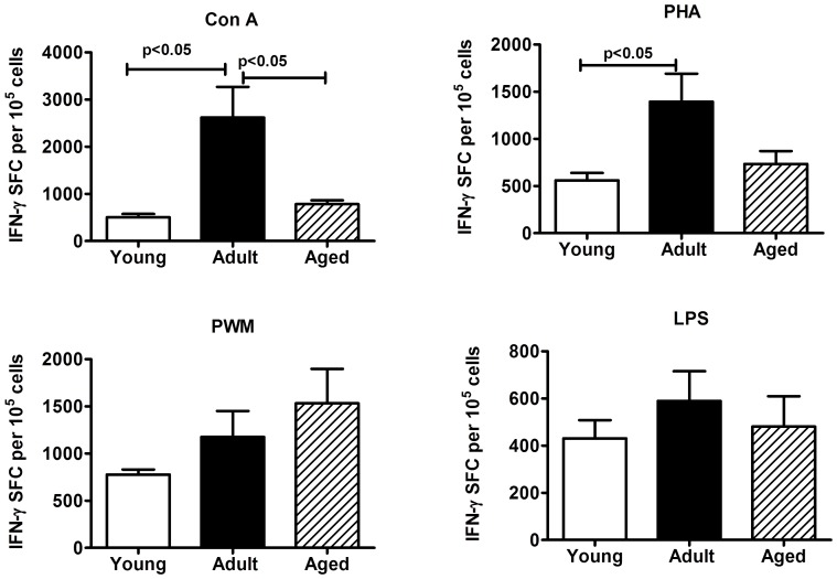
Figure 5. IFN-γ ELISpot response to mitogens.
Triplicate wells of the 96-well microtiter plates, pre-coated with IFN-γ antibody, were seeded with 105 PBMC from the monkeys in the three different age groups studied and stimulated with 5 µg of each of the mitogens for 36 h at 37°C followed by washing and staining with biotynylated second IFN-γ antibody. The total number of spot forming cells (SFC) in each of the mitogen-stimulated wells was counted and adjusted to control medium as background. See methods section for experimental details. The results shown are average of two separate experiments and the standard deviation values did not exceed 15% of the mean value. P values were considered at p<0.05.

