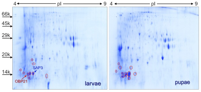
Figure 5. Two-dimensional gel electrophoretic separation of extracts from 100 fourth instar larvae and 100 pupae of An. gambiae.

The gel was stained with colloidal Coomassie Brilliant Blue and all the spots migrating with apparent molecular weight lower than 24(coverage by aminoacid sequence up to 61.87%), found in several spots (red circles). In larvae we could also detect OBP21 (Entry code in Uniprot Q8I8S3; coverage by aminoacid sequence 9.16%) and SAP3 (coverage by aminoacid sequence up to 18.25%), present in spots where also OBP9 was identified. Molecular weight markers are, from the top: Phosphorylase b, from rabbit muscle (97 kDa), Bovine serum albumin (66 kDa), Ovalbumin (45 kDa), Carbonic anhydrase (29 kDa), Trypsin inhibitor (20 kDa), α-Lactalbumin (14 kDa).
