Figure 2.
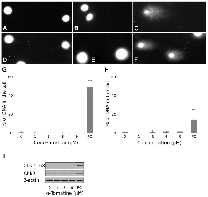
Detection of DNA damage. Digital camera image of the comet assay; the alkaline version (top row), (A) untreated MCF-7 cells, (B) 6 μM of α-tomatine-treated cells and (C) 1.5% H2O2-treated cells (positive control). Digital camera image of comet assay: the neutral version (bottom row), (D) untreated MCF-7 cells, (E) 6 μM of α-tomatine-treated cells and (F) cells after exposure of 20 Gy of irradiation (positive control). Microscope magnification, ×20. (G) The dependence of DNA single-strand breaks on the concentration of α-tomatine after 4 h of exposure in MCF-7 cells. Asterisks indicate values significantly (p<0.001) different from the control (distinguished using Student’s t-test). PC, positive control (1.5% H2O2). (H) The dependence of DNA double-strand breaks on the concentration of α-tomatine after 4 h of exposure in MCF-7 cells. Asterisk indicate values significantly (p<0.001) different from the control (Student’s t-test). PC, positive control (20 Gy γ radiation). (I) Changes in Chk2 and Chk2 phosphorylated on threonine 68 (Chk2_t68) detected by western blotting 4 h after exposure to 1, 3 and 6 μM of α-tomatine. To confirm equal protein loading, membranes were reincubated with β-actin. PC, positive control (10 Gy γ radiation after 24 h). Chk2, check-point kinase 2.

