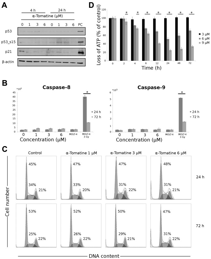
Figure 3.
The effect of α-tomatine on the induction of apoptosis in MCF-7 cells. (A) Changes in p53, p53 phosphorylated on serine 15 (p53_s15) and p21WAF1/Cip1 detected by western blotting 4 and 24 h after exposure to 1, 3 and 6 μM of α-tomatine. To confirm equal protein loading, membranes were reincubated with β-actin. PC, positive control (10 Gy γ radiation after 24 h). (B) The activity of caspase-8 and -9 was determined 24 and 72 h after exposure to 1, 3 and 6 μM of α-tomatine. γ irradiation (3 Gy) of MOLT-4 cells was used as a positive control. (C) Cell cycle distribution was measured using flow cytometric detection of DNA content in the cells. DNA content was analyzed 24 and 72 h after α-tomatine treatment in concentrations of 1, 3 and 6 μM. (D) The loss of ATP was measured using ATP bioluminescent assay kit at given time intervals after exposure to 3, 6 and 9 μM of α-tomatine. Values represent means ± SD of 3 independent experiments (*p≤0.05 compared with the untreated control group with one-way ANOVA test and Dunnett’s post-hoc test for multiple comparisons-loss of ATP and with Student’s t-test-activity of caspases and cell cycle distribution).

