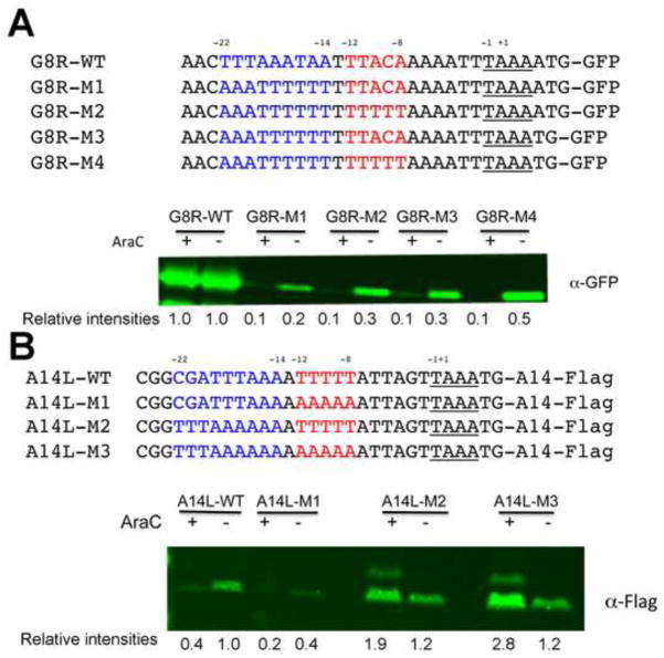
Fig. 4.
Mutational analyses of an intermediate and late promoter. (A) Intermediate promoter mutations. The sequences of the WT G8R promoter and mutated G8R promoters attached to GFP are shown. Plasmids containing these constructs were transfected into cells that had been infected with VACV in the absence (−) or presence (+) of AraC. After 16 h, the cells were lysed and analyzed by Western blotting with fluorescent antibody to GFP. Relative band intensities shown below the blot were determined using ImageJ. (B) Late promoter mutations. The WT A14L promoter and mutated A14L promoters were attached to the A14L ORF with a Flag tag. The sequences of the promoters are shown. Infection, transfection and Western blotting were similar to that of panel A except that antibody to the Flag epitope was used.

