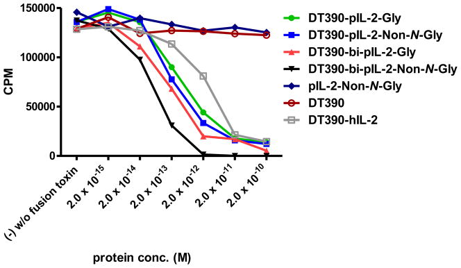
Figure 5.

In vitro Cell Proliferation Inhibition Analysis of the Four Porcine IL-2 Fusion Toxins Using LCL13271 Cells: 1) DT390-pIL-2-Gly (green line); 2) DT390-pIL-2-Non-N-Gly (blue line); 3) DT390-bi-pIL-2-Gly (red line); 4) DT390-bi-pIL-2-Non-N-Gly (black line); 5) pIL-2-Non-N-Gly alone (navy blue); 6) DT390 alone (pink); 7) Ontak®-like monovalent human IL-2 fusion toxin (DT390-hIL-2) (brown).Y-axis: cpm value by incorporating the tritium-labeled thymidine. X-axis: plated IL-2 fusion toxin concentration. Cyclohexmide (1:8) was used as a positive control. The negative control contained cells only without fusion toxin. Data are representative of three individual assays.
