
Figure 1. Fear extinction silences the basal amygdala fear memory circuit.
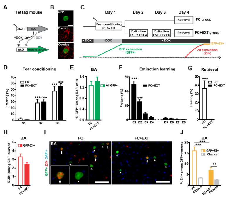
(A) A double transgenic TetTag mouse line was used that expresses the tetracycline transcriptional activator (tTA) under control of the activity-regulated c-fos promoter. In the absence of doxycycline (- DOX), tTA binds to the tet operator (tetO) in the second transgene and induces expression of a long-lasting histone2B-GFP fusion protein. (B) Image of an excitatory basal amygdala (BA) neuron tagged with long-lasting histone2B/GFP (green) and immunolabeled with a Calcium/calmodulin-dependent kinase II (CamKIIa) antibody (red). (C) Schema of the experimental procedure. “-DOX” opened a time-window for tagging fear conditioning activated neurons (GFP+; i.e. fear neurons) in two experimental groups (FC, n=15; FC+EXT, n=17). Zif expression during retrieval (Zif+) was used to detect the reactivation of tagged fear neurons (GFP+Zif+; i.e. active fear neurons). The absence of Zif was used to identify silent fear neurons (GFP+Zif-). (D) As training progressed, FC and FC+EXT mice showed an increase in their level of freezing (between sessions: p<0.001 *** for FC and °°° for FC+EXT). (E) FC and FC+EXT groups had similar percentages of GFP+ neurons (sum of GFP+Zif- and GFP+Zif+) in the BA (p=0.49). (F) The FC+EXT group showed a significant decrease in freezing during the extinction sessions on day 2 and 3 (p<0.001: *** E1 vs. others and °°° E2 vs. others). (G) Freezing during retrieval on day 4 in the FC+EXT group was absent and was significantly lower compared to the FC group (p=0.00014). (H) There was no significant difference in the percentage of Zif+ among GFP- neurons between the two groups (p=0.064). (I) Representative image of GFP-positive (GFP+, green) and Zif-positive (Zif+, red) nuclei in the basal amygdala (BA) of a FC (left) and FC+EXT mouse (right). Arrows indicate active fear neurons (GFP+Zif+) and arrowheads indicate silent fear neurons (GFP+Zif-). (J) Extinction decreased the number of active BA fear neurons, as indicated by FC+EXT mice having less GFP+Zif+ neurons than FC mice (p=0.00023). In both groups the percentage of GFP+ZIF+ neurons was higher than chance level (FC: p=0.0000098; FC+EXT p= 0.0014). Chance level was determined by using the % of Zif+ among GFP- neurons as shown in (H). Scale bar, 50 μm. Graphs show means ± SEM. **P < 0.01, ***P < 0.001.
See also Figure S1.