
Figure 2. Activation of brain regions upstream of the basal amygdala is not altered after fear extinction.
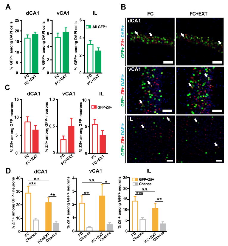
(A) The FC and FC+EXT groups had similar percentages of GFP+ neurons in the dCA1, vCA1 and IL (dCA1: p=0.44; vCA1: p=0.50; IL: p=0.17). (B) Representative image of GFP+ neurons (green) and Zif+ neurons (red) in the dorsal CA1 (dCA1, top), ventral CA1 (vCA1, middle) and infralimbic cortex (IL, bottom) of a FC mouse (left) and a FC+EXT mouse (right). Blue: DAPI. Arrows indicate active fear neurons (GFP+Zif+). Scale bar, 50 um. (C) No significant differences in the percentage of Zif+ among GFP- neurons were found (dCA1: p=0.25; vCA1: p=0.21; IL: p=0.056). (D) Extinction had no effect on reactivation of dCA1, vCA1 and IL. The number of GFP+Zif+ neurons was similar between the two groups (dCA1: p=0.26; vCA1: p=0.61; IL: p=0.27). The number of GFP+Zif+ neurons was above chance level in the dCA1, vCA1 and IL of both groups (dCA1: FC vs. chance, p=0.00017; FC-EXT vs. chance, p=0.000017; vCA1: FC vs. chance, p=0.0027; FC-EXT vs. chance, p=0.0072; IL: FC vs. chance, p=0.000062; FC-EXT vs. chance, p=0.0026). Chance level was determined by using the % of Zif+ among GFP- neurons as shown in (C). Graphs show means ± SEM. *P < 0.05, **P < 0.01, ***P < 0.001.