
Figure 4. Fear extinction increases perisomatic parvalbumin around silent BA fear neurons.
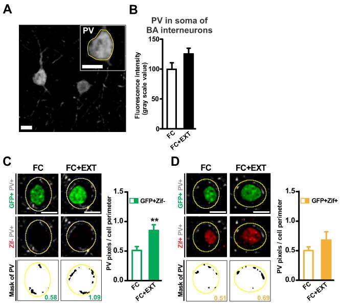
(A) Representative image of a parvalbumin-positive (PV+) interneuron soma in the basal amygdala (BA; scale bar: 10μm). (B) Extinction had no effect on somatic expression of PV (p=0.064). (C-D left) Representative images of perisomatic PV immunolabeling around silent BA fear neurons (C, GFP+Zif-) and active BA fear neurons (D, GFP+Zif+). Three images are shown for each fear neuron (top: GFP in green, PV in grey; middle: Zif in red, PV in grey; bottom: PV mask in black, value for that neuron obtained by dividing black pixels by perimeter of yellow outline). Scale bars, 10 μm. (C-D right) Extinction increased perisomatic PV around silent fear neurons (C, p=0.0061) but not around active fear neurons (D, p=0.18). Graphs show means ± s.e.m. **P < 0.01.
See also Figure S3.