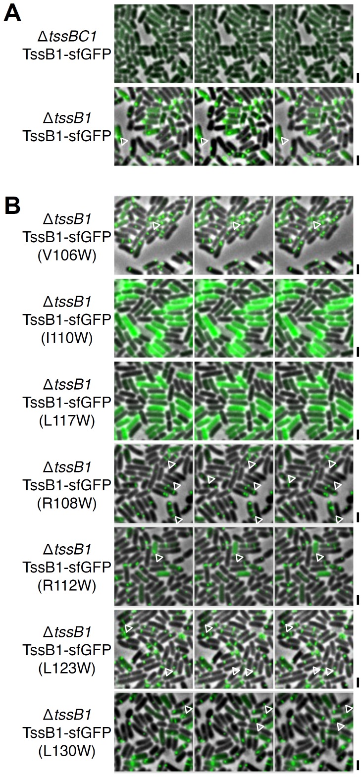
Figure 5. The hydrophobic motif of the TssB1 α-helix is required for T6SS sheath assembly.
Time-lapse fluorescence microscopy recordings showing sheath dynamics in ΔtssBC1 (ΔtssB1-ΔtssC1) or ΔtssB1 cells producing TssB1-sfGFP (TssB1) (A) or TssB1-sfGFP bearing the indicated substitutions (B). Individual images were taken every 30 sec. Assembly and contraction events are indicated by the white open triangles. Scale bars are 2 µm.

