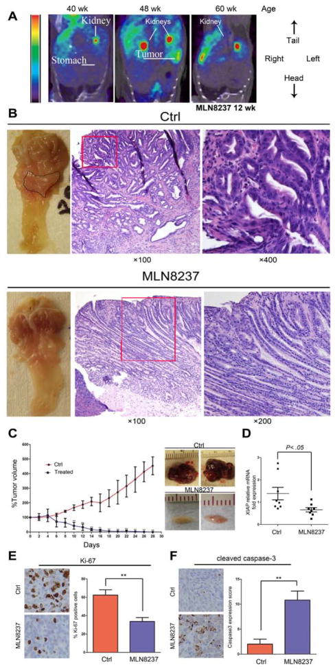
Figure 6. MLN8237 treatment reduces tumor growth in vivo.
A) A representative microPET coronal view of a mouse injected with [18F]FLT and imaged before and after the MLN8237 treatment. The tumors are invisible after 12 weeks post treatment. Because of the high uptake, kidneys showed typical [18F]FLT staining. B) Representative images of the antrum and H&E staining of gastric mucosa of Tff1-knockout mice. While tumors were visibly large in control (upper left panel), they became undetectable or small after 3 months of treatment (lower left panel). H&E staining indicated adenocarcinoma in control animals (upper middle and right panels) whereas MLN8237-treated animals exhibited low grade dysplasia (lower middle and right panels). C) AGS tumor xenografts were treated with MLN8237 (30 mg/kg) for 28 days. Tumor volume was dramatically reduced in the treated group as illustrated in the graph (p<0.01) (left panel). After collection of tumors at the end of experiment, representative tumors from the control or MLN8237 treated groups are shown (right panel). D) Real-time RT-PCR analysis of XIAP in xenograft tumors in control or ML8237- treated animals. E) Representative images of immunohistochemical analysis of Ki-67 protein expression in MLN8237-treated or control xenografts. F) Representative images of Immunohistochemical analysis of cleaved caspase-3 protein expression in MLN8237-treated or control xenografts.

