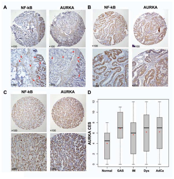
Figure 7. Expression of AURKA and NF-κB is associated with human gastric cancer development and progression.
A) IHC staining for AURKA and NF-κB on consecutive replicates of the same tissue sample. Normal gastric glands with negative AURKA and NF-κB immunostaining are indicated by arrow-heads whereas glands with intestinal metaplasia showing positive AURKA and NF-κB staining are indicated with arrows. B) IHC staining for AURKA and NF-κB on consecutive replicates of moderately differentiated gastric adenocarcinoma showing strong immunostaining for AURKA and NF-κB. C) The same as in panel B showing a case of poorly differentiated gastric adenocarcinoma. D) The graph summarizes the AURKA immunohistochemical staining results on gastric tissue microarrays. Horizontal bars indicate the median whereas red dots depict the mean. Normal, normal gastric glands; GAS, gastritis; IM, intestinal metaplasia; Dys, dysplasia; AdCa, adenocarcinoma. AURKA was significantly overexpressed in all stages of gastric tumorigenesis (p<0.001).

