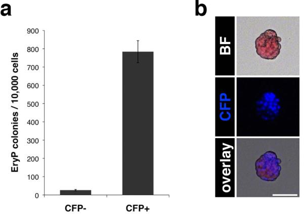
Figure 4. Nearly all EryP progenitor activity is present in the CFP positive population.
(a) Representative EryP progenitor assay of CFP-positive and -negative cells sorted from E8.5 ε-globin-H2B-CFP embryos (mean of 3 technical replicates ± SD). The experiment was repeated at least 7 times, with comparable results. (b) Bright field and fluorescent images of an EryP colony. Images were acquired using a Zeiss AxioCam color camera mounted on a Zeiss Axio Observer Z1 inverted microscope and outfitted with a EC Plan-Neofluar 10x/0.30 objective. Scale bar, 50 μm.

