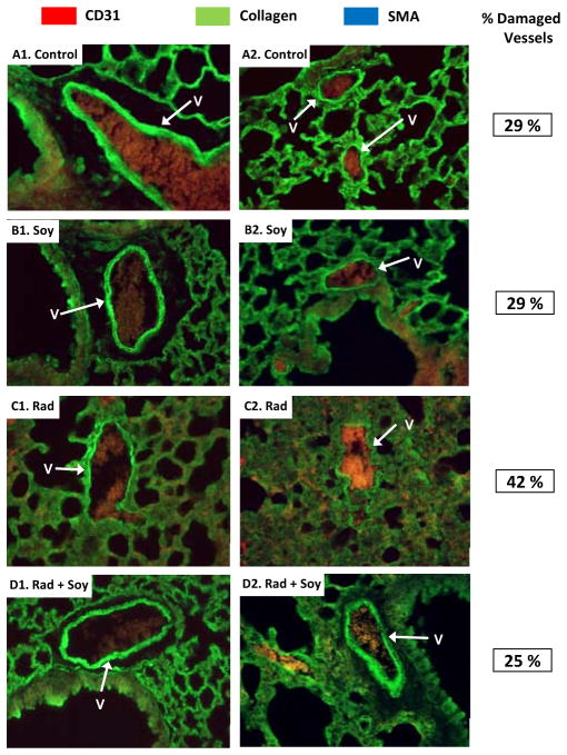
Fig 2. Fluorescent staining of vasculature in lung tissue sections.
Lung tissue sections were immunostained with fluorescent dyes, including red for endothelial cells (anti-CD31), blue for pericytes (anti-SMA) and green for collagen (anti-collagen IV) as detailed in Materials & Methods. Representative images of large and small vessels (white arrows) of the lung tissues are presented. (A1–2) Vessels with integral basement membranes from control mice. (B 1–2) Vessels from soy-treated mice showing regular green collagen staining of vessel basement membranes. (C1–2) Vessels from radiation-treated mice (Rad) showing disruption or distortion in the continuity of the collagen staining of basement membranes as well as thickening of alveolar septa surrounding the vessels (C2). (D1–2) Vessels from radiation and soy treated mice (Rad + Soy) showing integrity and continuity of vessel walls. The percentage of damaged vessels estimated in 20 fields of 40X, obtained from two mice per group, is reported in the third column. All magnifications 40X.

