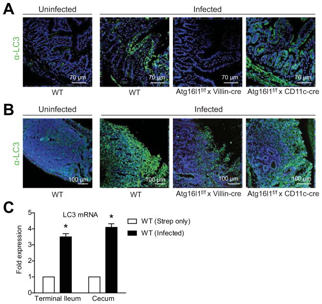
Figure 2. LC3 expression is induced following in vivo S Typhimurium infection.
(A–B) Mice were infected with S Typhimurium and tissues were harvested after 24 hours. Terminal ileum (A) and cecum (B) were immunostained with DAPI (blue) and anti-LC3 (green) and imaged by confocal microscopy. Uninfected wild-type tissues are shown on the left. (C) LC3B mRNA levels from isolated wild-type ileal and cecal epithelial cells were determined by qPCR. Numbers shown are relative to streptomycin-only-treated wild-type mice. Bars indicate mean plus S.D. *P ≤ .01. Data are representative of at least 3 independent experiments (n = 9).

