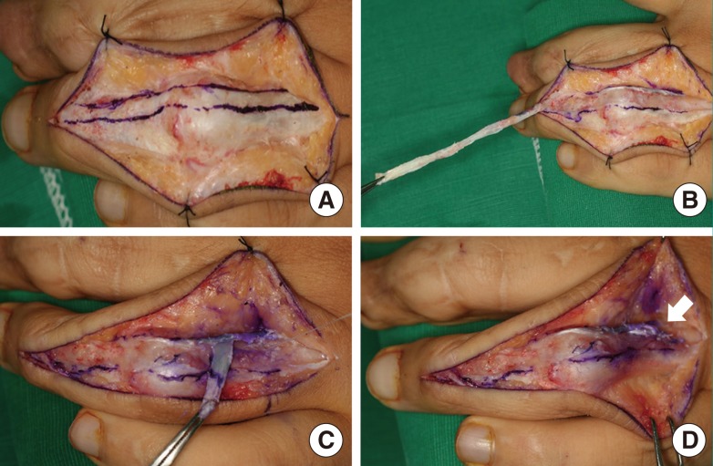
Fig. 1.
Intraoperative photograph of lateral band technique (group A)
(A) A midline longitudinal dorsal incision exposes the terminal extensor mechanism. (B) A healthy portion of the lateral band is mobilized for reconstruction of the oblique retinacular ligament. (C) The band is re-routed through a deep volar tunnel (not visible) and retrieved proximally on the contralateral side. (D) The lateral band is secured to the proximal phalanx (white arrow).

