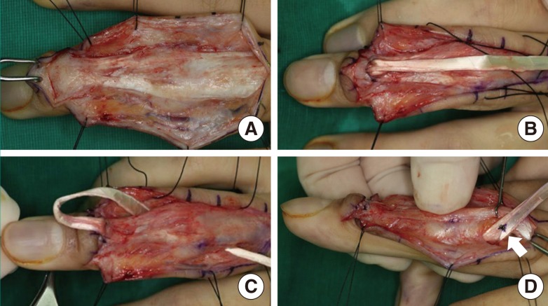
Fig. 2.
Intraoperative photograph of free tendon technique (group B)
(A) The same longitudinal incision is used for free tendon ORL reconstruction. In this patient, the lateral band is attenuated and cannot be used for ORL reconstruction. (B) The distal end of a palmaris longus graft tendon is secured to the base of the distal phalanx. (C) The proximal end is routed through the volar tunnel. (D) Graft tension adjustment. The arrow points to the distal end of the graft to be secured to the proximal phalanx. ORL, oblique retinacular ligament.

