
Fig. 1.
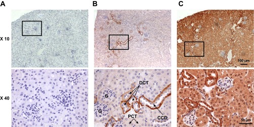
Immunohistochemical staining of Kir5.1 and Kir4.1 channels in the mouse kidney. Representative immunohistochemically stained images of kidney sections of C57BL/6 mice are shown at ×10 and ×40 magnifications. A: negative control tissue stained with secondary antibodies in the absence of primary antibodies. B and C: representative immunohistochemical staining for Kir5.1 (B) and Kir4.1 (C) channels. The boxes at ×10 optical image represent the magnified areas shown at ×40 optical magnification. Scale bars are shown. G, glomerulus; PCT, proximal convoluted tubule; DCT, distal convoluted tubule; CCD, cortical collecting duct.