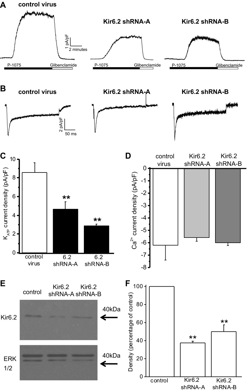
Fig. 1.
Short hairpin (sh)RNA knockdown of ATP-sensitive K+ (KATP) currents in cardiac myocytes. A: whole cell recordings of KATP currents from isolated ventricular myocytes after 24-h infection with adenovirus expressing either control shRNA, Kir6.2 shRNA-A, or Kir6.2 shRNA-B sequences. Currents were recorded at 0 mV, and P1075 (10 μM) and glibenclamide (10 μM) were bath applied, as indicated by the horizontal bars. B: whole cell voltage-clamp recordings of voltage-gated Ca2+ currents from single ventricular myocytes after 24-h infection with either control shRNA, Kir6.2 shRNA-A, or Kir6.2 shRNA-B adenoviruses. Ca2+ currents were evoked by a 240-ms voltage step from −40 to 0 mV. C: peak KATP currents normalized for cell size (in pA/pF) of myocytes infected with control shRNA (n = 17), Kir6.2 shRNA-A (n = 12), or Kir6.2 shRNA-B (n = 5). **P < 0.01 vs. control. D: mean peak Ca2+ currents normalized for myocyte size (in pA/pF) recorded from cardiomyocytes infected with either control shRNA, Kir6.2 shRNA-A, or Kir6.2 shRNA-B adenoviruses (n = 3 for each). E: representative Western blots showing Kir6.2 protein from isolated ventricular myocytes after 24-h infection expressing control shRNA, Kir6.2 shRNA-A, or Kir6.2 shRNA-B. Total ERK was used as a loading control. F: average densitometry showing the effects of Kir6.2 shRNA on Kir6.2 protein levels. n = 3 Western blots from 3 hearts. **P < 0.01. Stats were done on raw densitometry values.

