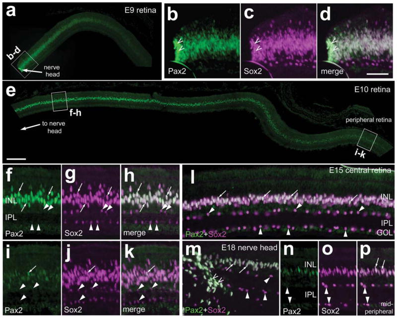
Figure 1.
Pax2 is expressed in the chick retina during late stages of embryonic development. Vertical sections of retinas from E9 (a–d), E10 (e–k), E15 (l), and E18 (m–p) chick embryos were labeled with antibodies to Pax2 and Sox2. b–d, f–h, and i–k are threefold enlargements of the boxed areas in a and e. m: Micrograph of the optic nerve head and retina from an E18 eye. n–p are micrographs of the midperipheral retina about 2 mm peripheral to the optic nerve in m. Arrowheads indicate cholinergic amacrine cells labeled for Sox2 alone; arrows indicate cells labeled for Sox2 and Pax2; carets indicate peripapillary cells. INL, inner nuclear layer; IPL, inner plexiform layer; GCL, ganglion cell layer. Scale bars = 50 μm in e (applies to a,e); 50 μm in d (applies b–d,f–p). [Color figure can be viewed in the online issue, which is available at www.interscience.wiley.com.]

