Figure 5.
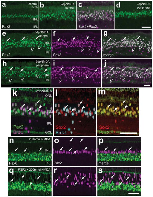
a–s: Pax2 is expressed by Müller glia that reenter the cell cycle in acutely damaged retinas. Vertical sections of chick retina were labeled with antibodies to Sox2 (magenta in a–I; red in l and m), Pax2 (green in a–m; magenta in n–s), BrdU (magenta in k; turquoise in l), and Pax6 (green in n–s). Retinas were obtained from eyes that were injected with saline (a), 2,000 nmol NMDA (b–m), or 200 ng FGF2 at P5 and P6 and 200 nmol NMDA at P7 (n–s). Retinas were harvested 6 hours after injection of BrdU at 2 (b–d and k–m) and 3 (e–j) days after NMDA treatment or 24 hours after injection of 200 nmol NMDA with FGF2 pretreatment (n–s). Widefield (a–m) and confocal (n–s) microscopy were used to obtain images. Arrows indicate nuclei of Müller glia that are labeled for Pax2 and BrdU, Sox2, or Pax6; small double arrows indicate the nuclei of presumptive NIRG cells that are labeled for Sox2 alone (e–j); arrowheads indicate nuclei of Müller glia that are labeled for Pax2 and Sox2 but not BrdU (k–m). INL, inner nuclear layer; IPL, inner plexiform layer; GCL, ganglion cell layer. Scale bars = 50 μm in d (applies to a–d); 50 μm in j (applies to e–j); 50 μm in m (applies to k–m); 50 μm in s (applies to o–s). [Color figure can be viewed in the online issue, which is available at www.interscience.wiley.com.]

