Figure 8.
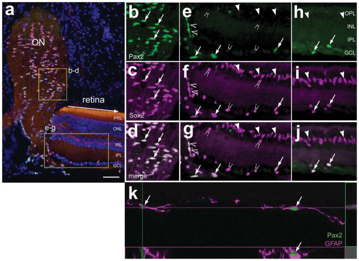
a–k: In the mouse eye, Pax2 is expressed by cells in the optic nerve head and retinal astrocytes. Transverse sections of the eye were labeled with DAPI (blue in a,j) and antibodies to Pax2 (green), Sox2 (magenta in a–j), and GFAP (magenta in k). Images were obtained using widefield (a–j) and confocal (k) microscopy. b–g: Twofold enlargements of the boxed areas in a. Arrowheads indicate Sox2+ nuclei of Müller glia; carets indicate Sox2+/Pax2+ peripapillary glia; small double arrows indicate the Sox2+ nuclei of cholinergic amacrine cells; arrows indicate astrocytes in the GCL or NFL in the retina; k includes orthogonal projections. ONL, outer nuclear layer; INL, inner nuclear layer; IPL, inner plexiform layer; GCL, ganglion cell layer; NFL, nerve fiber layer. Scale bar = 50 μm for a. [Color figure can be viewed in the online issue, which is available at www.interscience.wiley.com.]

