
Figure 5. Rescue of TDP-43 in motor neurons.
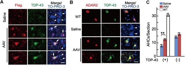
- Immunohistochemistry for TDP-43 (green) and Flag (red) in the spinal cord. AHCs were recognized as large (≥20 µm diameter) and TO-PRO-3-positive (blue) cells in the AH. Flag-positive AHCs exhibited intense nuclear and faint cytoplasmic TDP-43 immunoreactivity (arrows) in AAV9-injected AR2 mice (AAV). Arrowheads indicate AHCs negative for both Flag immunoreactivity and TDP-43 immunoreactivity in the nucleus in saline-injected AR2 mice (Saline) and AAV. TO-PRO-3 was used as a cell marker. Scale bar is 20 μm.
- Double immunostaining of the AH for TDP-43 and ADAR2. The nuclei of the AHCs were either double-positive or double-negative for TDP-43 (green) and ADAR2 (red) in AAV, Saline and wild-type mice (WT). Scale bar is 20 μm.
- Number of TDP-43-positive and -negative AHCs in Saline (blue columns), AAV (red columns) and WT (white column) (n = 5 for each group). All error bars represent the s.e.m. **p < 0.01 (Student's t-test).