
Figure 1. Oral administration of ALK5 inhibitors suppresses melanoma and LN metastases with enhanced CTL activity.
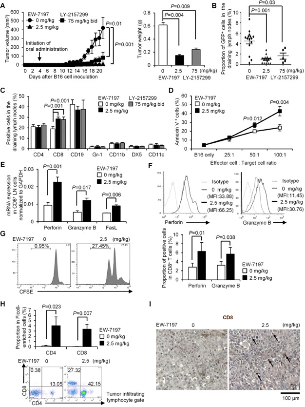
C57BL/6 mice were treated with vehicle or EW-7197 (2.5 mg/kg daily) (n = 15/group)/LY-2157299 (75 mg/kg bid) (n = 5) from 4 days after inoculation of GFP-expressing B16 cells (4 × 10⁴) into the left footpads. Data are shown as mean ± SEM. P values were calculated by 2-tailed unpaired Student's t-test or by two-way ANOVA test for (A).
- A. Chronological tumour volumes (left), tumour weights on Day 21 (right).
- B,C. The % of GFP+ B16 cells (medians ± interquartile) and immune cell subsets in dLNs were determined by flowcytometry.
- D. Target cytolysis at the indicated ratios of effector CD8+ T cells: target B16 cells was evaluated by annexin V/PI.
- E. qPCR analyses for mRNA levels of the cytolytic molecules in CD8+ dLN cells (n = 5/group).
- F. Histograms show CD8+ gate with MFI. Graphs show the % of positive cells in CD8+ gate (n = 10/group).
- G. Proliferation of CD8+ dLN cells stimulated with gp100 peptide was assessed by CFSE dilution.
- H. Representative CD4/8 dot plots of TILs. Graphs show the % of CD4+ or CD8+ cells in the Ficoll-enriched cells (n = 8/group).
- I. Representative immunohistochemistry sections of inoculated melanomas (scale bar: 100 μm). Arrows indicate CD8+ cells.