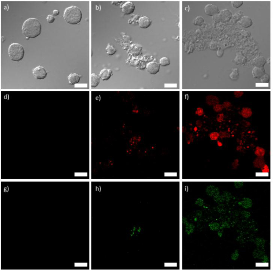
Figure 3.
a–c) Differential interference contrast (DIC) images and d–i) fluorescence images of Jurkat cells incubated with 3a (d,g), 3b (e,h), and 3c (f,i). Red fluorescence (d–f) is from the Ru(bpy)32+-doped particles and green fluorescence (g–i) is from the Annexin V FITC conjugate early apoptosis stain. Scale bars represent 25 µm.

