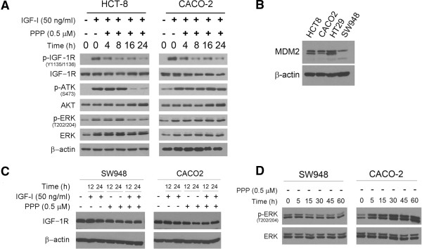
Figure 2.

PPP treatment triggers apoptosis in TP53 wild-type but not mutated cells. (A). The TP53 wild-type HCT-8 and mutated CACO-2 cells were treated with 500 nM PPP in the presence or absence of 50 ng/ml IGF-I for the hours as indicated. The treated cells were then examined by western blotting for the presence of the phosphorylated and unphosphorylated IGF-1R, AKT and ERK with β-actin as the loading control. (B). The TP53 wild-type HCT8 and SW948 and mutated CACO-2 and HT29 were examined by western blotting for the levels of MDM2 protein. (C). The TP53 wild-type SW948 and mutated CACO-2 cells were treated with 500 nM PPP and 50 ng/ml IGF-I, alone and in combination, for the indicated hours. The cells were then examined by western blotting for the levels of IGF-1R protein. (D). SW948 and CACO-2 cells were treated with 500 nM PPP for the indicated minutes and then analyzed by western blotting for the levels of unphosphorylated and phosphorylated ERK (p-ERK).
