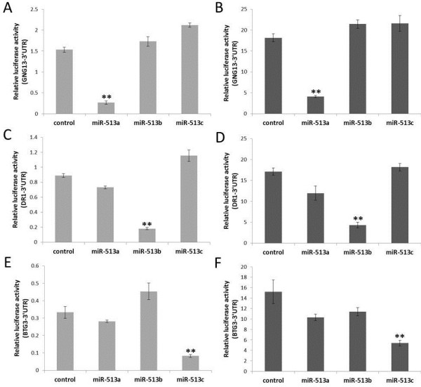
Figure 4.

Identification of miR-513 binding sites by luciferase assays. The Y-axis represents the ratio of Renilla luciferase (with candidate target gene’s 3′ UTR) activity to firefly luciferase activity after treated with negative control or miR-513 mimics. (A) and (B) show miR-513a’s specific targeting of predicted sites in the 3′UTR of GNG13. (C) and (D) indicate miR-513b’s specific targeting of the 3′UTR of DR1. (E) and (F) display miR-513c’sspecific targeting of the 3′UTR of BTG3. A, C, E are results in HEK293T cells, while B, D, F are results in HeLa cells. Error bars represent standard deviations (n = 3). ** P < 0.01 (two-tailed student’s t-test).
