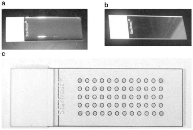
Fig. 2.
Images of agarose arrays during the preparation and selection of bound members. (a) An agarose slide that has solidified to a gel. (b) An agarose-coated microarray that has dried to a clear film. (c) An image of an agarose array that is placed on top of a paper grid to image use as a guide for spotting ligands; the spots to indicate the four corners of the grid have been put on the array using a hydrophobic marker and help align the grid for precise excision of bound nucleic acid on an array. The same grid is used to align the image of slides after hybridization with an RNA library.

