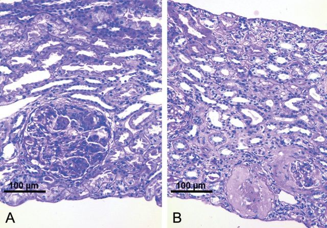
Fig. 1.
Renal biopsy findings in a 60-year-old patient with pauci-immune extracapillary-proliferative glomerulonephritis due to IgG kappa multiple myeloma. (A) Biopsy before bortezomib treatment (200×, PAS) showing a proliferating glomerular crescent. The tubulo-interstitial space appears to be normal. Polymorphonuclear leucocytes within the glomerular tuft. No infiltrates. (B) Biopsy after bortezomib treatment (200×, PAS) showing sclerosing glomeruli and sclerosing crescents, interstitial fibrosis and tubular atrophy with only scarce inflammatory infiltrates. The majority of glomeruli showed normal appearance.

