Figure.
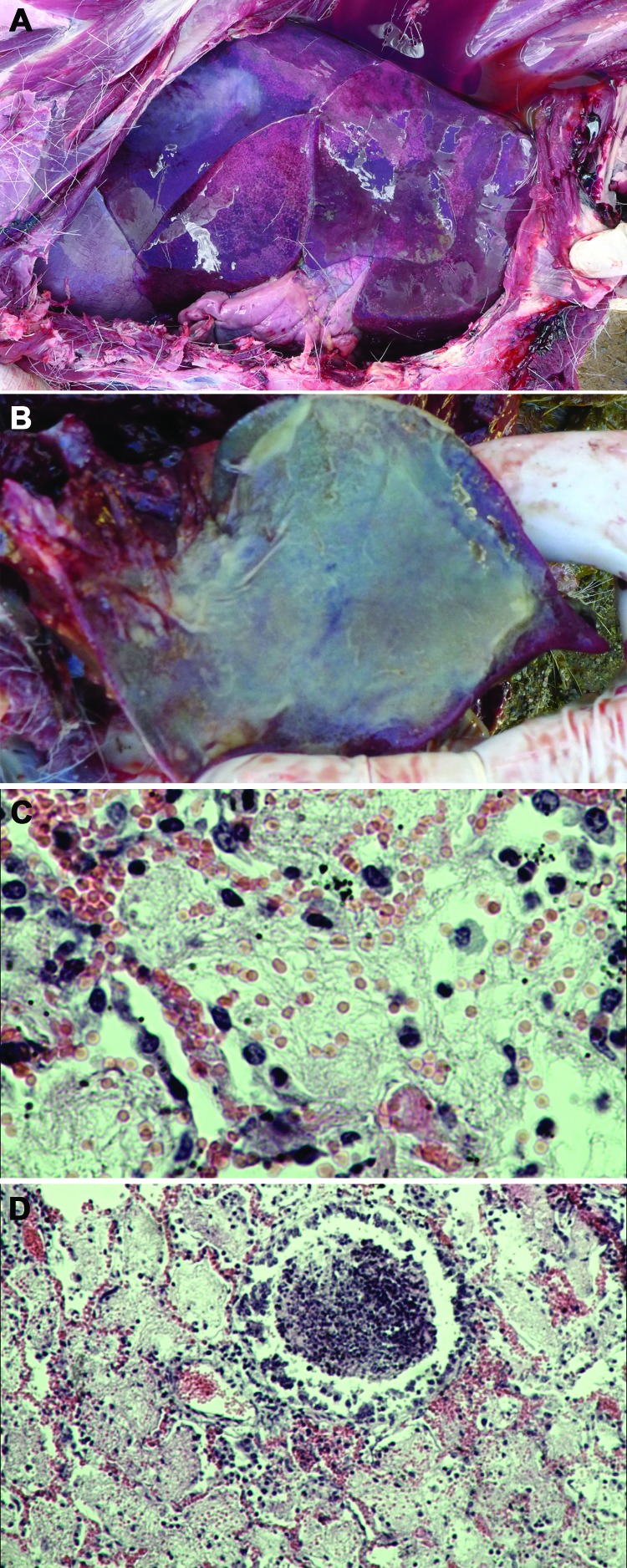
Pneumonia caused by Mycoplasma capricolum subsp. capripneumoniae in Tibetan antelope (Pantholops hodgsonii), Tibet, 2012. A) Lung of a caprine pleuropneumonia–infected Tibetan antelope (sample SZM2) showing lung hepatization. B) Lung of a caprine pleuropneumonia–infected Tibetan antelope (sample SH3) showing fibrin deposition. C and D) Fibrinous pneumonia with serofibrinous fluid and an inflammatory cell infiltrate, consisting of mainly lymphocytes, in the alveoli (panel C, sample SZM2, hematoxylin and eosin stain; original magnification ×400) and bronchioles (panel D, sample SH3, hematoxylin and eosin stain; original magnification ×100). Refer to Technical Appendix Table 1 for details of the lung samples used to generate images for this figure.

