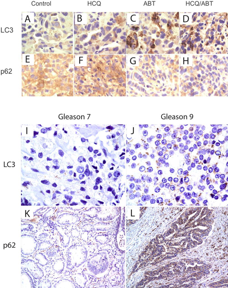
Figure 4.

Assessment of LC-3 and p62 in human tumor xenograft and human tissue. 4A-H: Histologic sections were obtained from the tumors of the mice in each treatment group from xenograft studies. LC3 and p62 antibody staining was performed in the tumor tissue sections from Control (A and E), ABT-737 (C and G), HCQ (B and F) and combination of ABT-737 and HCQ (D and H) by immunohistochemistry at 14 days of treatment. 4I–L: Assessment of LC3 and p62 in human cancers with low (Gleason Score 7) and high (Gleason Score 9) grade tumors.
