
Figure 3.
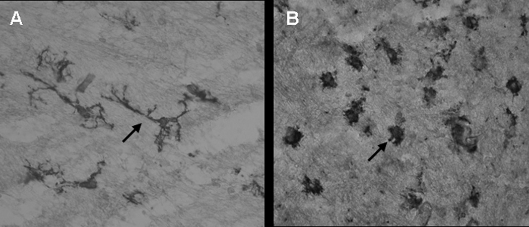
a. Tomato lectin staining of microglial cells in postnatal day 1 brain of rabbit kits born to dams injected intrauterine with saline (control) (A) and 20ug/kg LPS (endotoxin) (B).
Microglia in the control group exhibits ramified morphology (A, arrow) whereas in the endotoxin group the processess are shorter and thicker(B ,arrow). An increase in the density of amoeboid microglial cells in the endotoxin treated group was noted when compared to the control group.
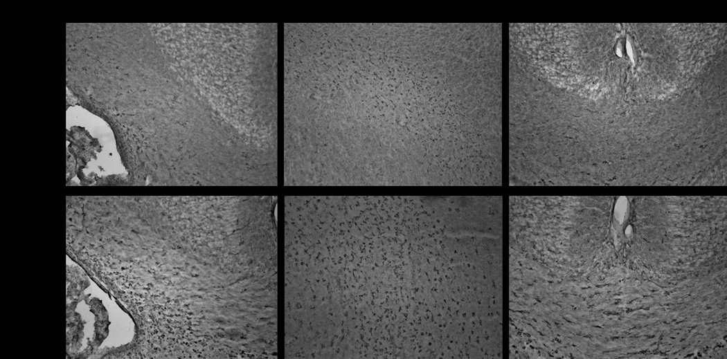
b. Tomato lectin staining of microglia in postnatal day1 control and endotoxin exposed kits.
Representative images indicate a robust increase in microglial cells along the border of lateral ventricle, internal capsule and corpus callosum in the endotoxin kit when compared to control.