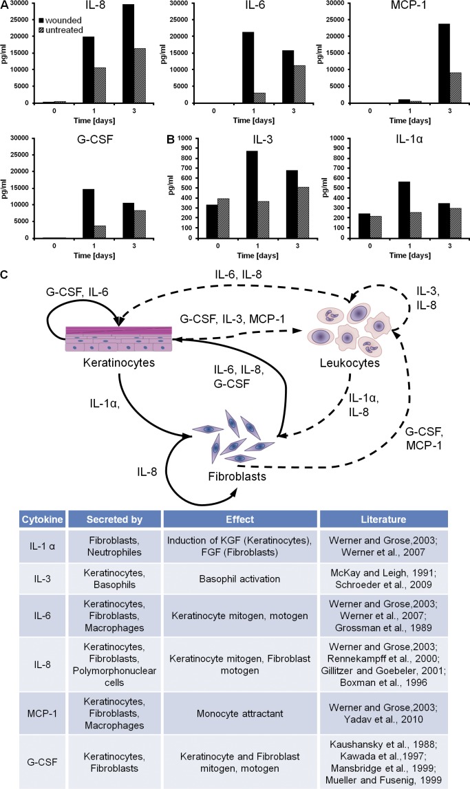
Figure 2.
Paracrine signaling of keratinocytes and fibroblasts in the excisional 3D in vitro wound-healing model. (A and B) Mean concentrations of growth factors measured in the supernatant of the 3D in vitro wound-healing model. The growth factors are divided into two groups, based on the maximum mean concentration: >10,000 pg/ml (A); <1,000 pg/ml (B). (C) The paracrine signaling of keratinocytes and fibroblasts under wound-healing conditions is shown. Arrows mark dependencies between the cell types. Dashed lines represent possible dependencies, which could not be measured in the 3D in vitro wound-healing model caused by the lack of leukocytes. Growth factor–secreting cell types and the impact of the factors on cell behavior are listed (Kaushansky et al., 1988; Grossman et al., 1989; Demetri and Griffin, 1991; McKay and Leigh, 1991; Boxman et al., 1996; Kawada et al., 1997; Mansbridge et al., 1999; Mueller and Fusenig, 1999; Rennekampff et al., 2000; Gillitzer and Goebeler, 2001; Angel and Szabowski, 2002; Werner and Grose, 2003; Schroeder et al., 2009; Yadav et al., 2010). Data are means; n = 4 from a single experiment.

