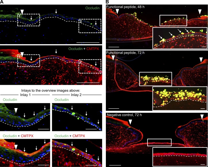
Figure 9.
Tight junction perturbation using a synthetic peptide emulating occludin loop B. (A) Histological sections of representative EETs showing occludin CMTPX colocalization (white arrows) in the suprabasal compartment over the full length of the EET. (B) A synthetic functional peptide emulating occludin loop B was used to block tight junction assembly during tongue extension. The integration of the biotin-tagged peptide into the EET was detected by applying streptavidin-FITC (green). Occludin-blocked cells failed to integrate in the ESM and were shed during tongue extension (white arrows). Insets show magnified areas of the EET to illustrate the effect of the functional peptide as well as the effect of the negative control. Arrowheads indicate the wound margin. Broken lines denote dermal–epidermal junction. Bars: (overview screens) 250 µm; (inlays and insets) 50 µm.

