Figure 1.
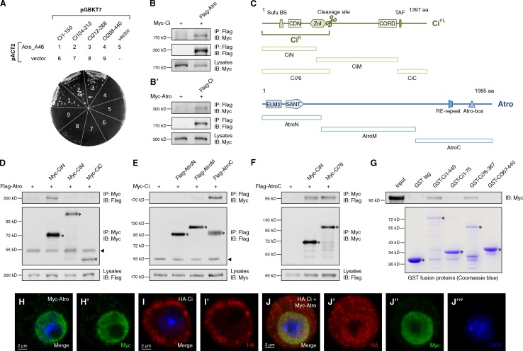
AtroC binds to the CiN. (A) Yeast two-hybrid assay between Atro_A46 fragment and indicated Ci fragments. (B and B′) Western blots of immunoprecipitates (top two panels) or lysates (bottom) from S2 cells expressing the indicated proteins. (C) Schematic representation of domains and motifs in Atro and Ci proteins and their fragments used in subsequent coIP assay. (D–F) Western blots of immunoprecipitates (top two panels) or lysates (bottom) from S2 cells expressing the indicated proteins. (G) GST pull-down between Myc-tagged AtroC and GST or GST-tagged Ci fragments. (H–J‴) S2 cells expressing the indicated proteins were immunostained with HA (red), Myc (green) antibodies, and DAPI (blue) to visualize nuclei. In all blots, asterisks indicate the target proteins and arrowheads indicate IgG.

