Figure 3.
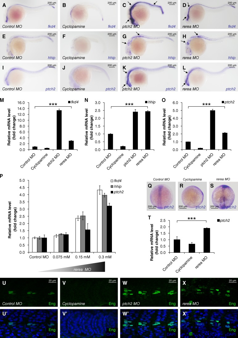
The repressor function of Atro in Hh signaling is evolutionally conserved in zebrafish. (A–L) Expression of Hh-responsive genes in zebrafish embryos that were injected with the indicated MOs or treated with cyclopamine (10 µM) at 24 hpf. Up-regulated in situ stainings are marked by arrows. (M–O) Relative mRNA levels of fkd4, hhip, and ptch2 from 24 hpf zebrafish embryos indicated in A–L were revealed by real-time PCR (mean ± SD; n ≥ 3). (P) Relative mRNA levels of fkd4, hhip, and ptch2 from 24 hpf zebrafish embryos, which were injected with gradient concentrations of rerea MO. (Q–S) Expression of ptch2 in zebrafish embryos injected with the indicated MOs or treated with cyclopamine (100 µM) at bud stage (10 hpf). (T) Relative mRNA levels of ptch2 from bud stage embryos indicated in Q–S were revealed by real-time PCR (mean ± SD; n ≥ 3). (U–X’) Zebrafish embryos injected with the indicated MOs or treated with cyclopamine (10 µM) at 24 hpf were immunostained with Eng antibody (green) and DAPI (blue) to visualize nuclei. P-values in this figure were obtained by student’s t test between two groups (***, P < 0.001).

