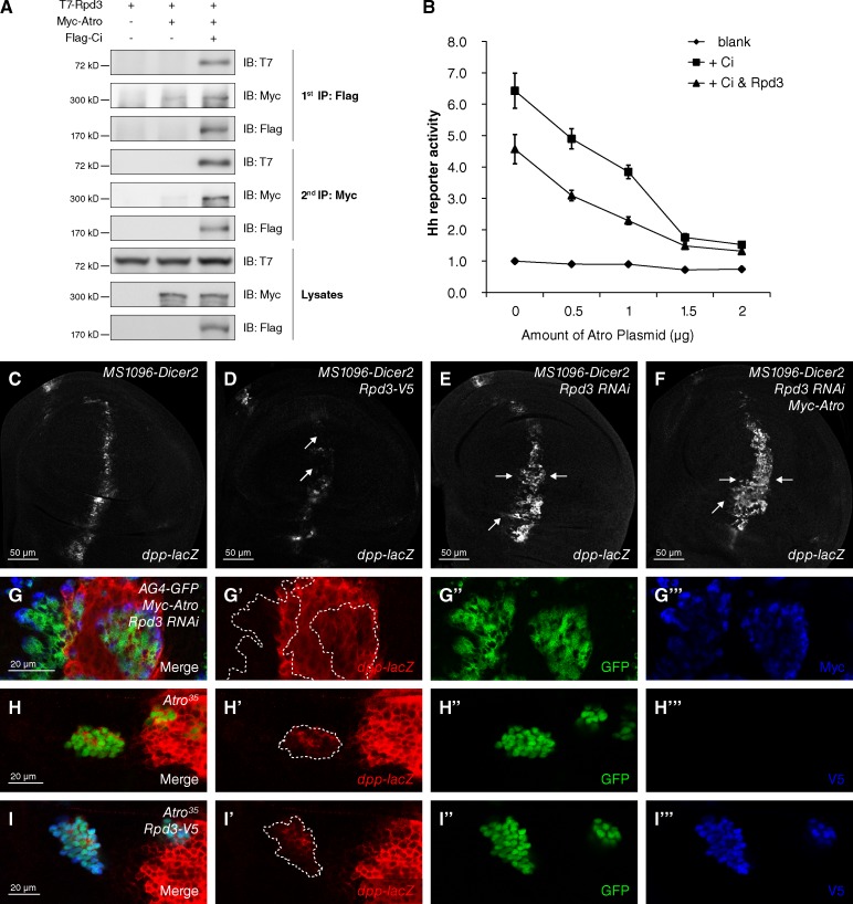
Figure 4.
Atro recruits Rpd3 to repress Hh signaling. (A) S2 cells expressing the indicated proteins were harvested for the two-step immunoprecipitation and analyzed by Western blotting. (B) Rpd3 cooperates with Atro to repress Hh signaling in reporter assay in S2 cells (mean ± SD; n = 3). The whole DNA amount transfected in each group was normalized equal with blank vectors. (C–F) A wild-type wing disc (C) or wing discs expressing Rpd3-V5 (D), Rpd3 RNAi (E), or Rpd3 RNAi plus Myc-Atro (F) with MS1096-Dicer2 Gal4 were immunostained to show the expression of dpp-lacZ. The level of dpp-lacZ was reduced in Rpd3 overexpressing disc (D, arrows) and increased in Rpd3 knockdown disc (E, arrows). Atro overexpression failed to block the up-regulated expression of dpp-lacZ by Rpd3 knockdown (F, arrows). (G–G‴) A wing disc expressing Myc-Atro plus Rpd3 RNAi with AG4-GFP was immunostained to show the expression of dpp-lacZ– (red), GFP- (green), and Myc-tagged Atro (blue). (H–I‴) Wing discs carrying Atro35 clones or Atro35 plus Rpd3 overexpression were immunostained to show the expression of dpp-lacZ (red) and GFP (green). From G to I‴, clones were marked by GFP-positive cells (dashed lines).

