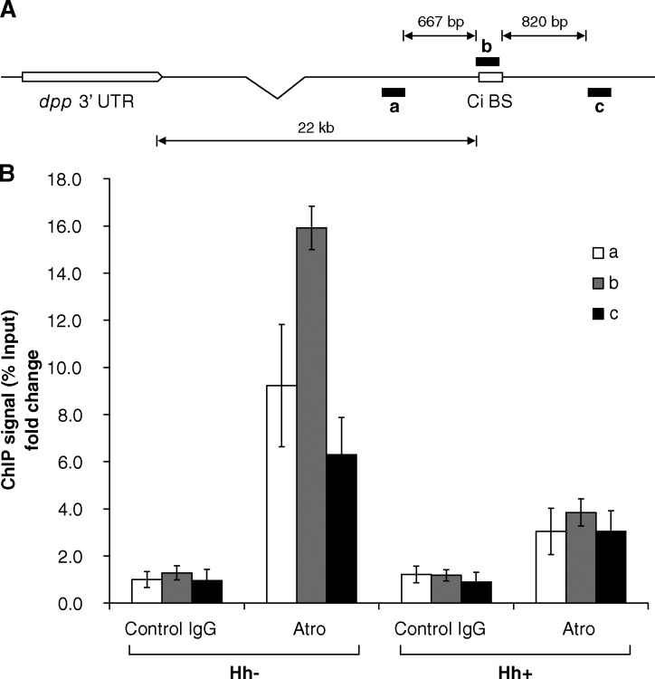
Figure 5.
Atro–Rpd3 complex associates with a Ci-binding site at the dpp locus in the absence of Hh signaling. (A) Schematic diagram of dpp locus and regions amplified with corresponding PCR primers to detect ChIP products. (B) ChIP for Atro around Ci binding site in dpp locus in Cl.8 cells with/without Hh treatment. Data of ChIP signals were normalized to 1/10 of input and showed as the fold change to the first group (mean ± SD; n = 3).

