. 2011 Jun;4(1):43–49. doi: 10.15283/ijsc.2011.4.1.43

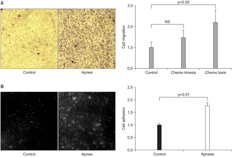
Fig. 4. (A) Examples of a field of view of transwell membrane showing stained mesenchymal stem cells (MSC) that migrated to the lower membrane side of the transwell when the lower compartment contained control serum (CONTROL) and serum from rats subjected to recurrent obstructive apneas (APNEA). Serum of control rats was placed in the lower transwell compartment in both cases. Normalized indices for the migration of MSC (chemotaxis and chemokinesis) induced by the serum of control and apneic rats are represented on the right side of the figure. Data are mean±SEM. NS: non-significant (p<0.05). (B) Examples of fields of view of the fluorescence-labelled MSC adhered to cultured endothelial cells when incubated in control serum (CONTROL) or in serum from rats subjected to recurrent obstructive apneas (APNEA). The normalized indices for the adhesion of MSC to endothelial cells in control rat serum and in serum from rats subjected to recurrent obstructive apneas are represented on the right panel. Data are mean±SEM. Reproduced from reference 24 with permission.

Fig. 4.